
Noncontrast vs. Contrast CT for USMLE Step 3 CCS
Which CT should you order for a suspected stroke, kidney stone, inflamed abdomen, or pulmonary embolism? Match the protocol to the clinical question.
On USMLE Step 3 CCS, the useful decision is more specific than “order a CT.” Choose the body region and a protocol that can answer the question in front of you. Noncontrast CT, routine IV contrast CT, and CT angiography (CTA) serve different purposes.
In this article
Noncontrast vs. contrast CT: Quick comparison
| Clinical question | Study to consider | Purpose |
|---|---|---|
| Suspected intracranial hemorrhage or acute stroke | CT head without IV contrast | Assess for bleeding promptly; add vascular imaging when indicated |
| Suspected appendicitis or diverticulitis in an adult | CT abdomen and pelvis with IV contrast | Evaluate inflammation and complications |
| Suspected acute mesenteric ischemia | CTA abdomen and pelvis with IV contrast | Evaluate mesenteric vessels and bowel |
| Suspected urinary stone when CT is needed | Low-dose CT abdomen and pelvis without IV contrast | Locate a stone and assess obstruction |
| Suspected pulmonary embolism (PE) when imaging is indicated | CT pulmonary angiography (CTPA) with IV contrast | Evaluate the pulmonary arteries |
These are common approaches, not universal orders. Clinical stability, pregnancy, kidney function, contrast history, and the possibility of a different diagnosis can change the plan.
CT head: Noncontrast first for suspected stroke
For a sudden focal neurologic deficit, CT head without IV contrast is commonly the initial brain imaging study. Its immediate role is to identify intracranial hemorrhage and other findings that affect treatment decisions. Establish the time of symptom onset and activate the appropriate acute stroke pathway while arranging imaging.
If a large-vessel occlusion is suspected, CTA of the head and neck may be added. MRI can detect some early or small infarcts more readily, but the first study depends on the stroke pathway and available resources. ACR: Cerebrovascular Disease
Noncontrast CT is also important for suspected intracranial injury when head imaging is indicated after trauma. Trauma alone does not mean every patient needs a scan. ACR: Head Trauma
CCS takeaway: In suspected acute stroke, order the initial noncontrast head CT promptly, then let the findings and stroke pathway guide further vascular imaging and treatment.
CT abdomen and pelvis: Contrast for inflammation, CTA for ischemia
Appendicitis and diverticulitis
When CT is the chosen test for an adult with suspected appendicitis or diverticulitis, CT abdomen and pelvis with IV contrast is usually appropriate. It helps define inflammation and complications, including collections. Oral contrast is not automatically required; the protocol varies with the diagnostic question and local practice. ACR: Suspected Appendicitis; ACR: Suspected Diverticulitis
Acute mesenteric ischemia
For suspected acute mesenteric ischemia, request CTA of the abdomen and pelvis with IV contrast. A dedicated angiographic protocol evaluates the mesenteric vessels; “CT abdomen with contrast” does not specify the same study. This is a time-sensitive concern that also needs urgent clinical assessment and specialty involvement. ACR: Imaging of Mesenteric Ischemia
CCS takeaway: Inflammatory disease and vascular ischemia may both call for IV contrast, but they can require different CT protocols.
Renal colic: A noncontrast stone protocol
When CT is needed to assess a suspected urinary stone, low-dose CT abdomen and pelvis without IV contrast is a common choice. It can show stone size and location as well as evidence of obstruction. Specify the noncontrast stone protocol in the order. ACR: Acute Onset Flank Pain—Suspicion of Stone Disease
It is not the right reflex for every patient with flank pain. Ultrasound is generally preferred initially during pregnancy, and another suspected emergency may require a different imaging approach. ACR: Stone Disease, Pregnancy Variant
CT chest: Use CTPA to evaluate pulmonary embolism
Assess clinical probability before ordering imaging for suspected PE. A D-dimer can help decide whether imaging is needed in appropriate patients with low or intermediate probability. When PE imaging is indicated and CT is suitable, order CT pulmonary angiography (CTPA) with IV contrast. Its contrast timing is designed to evaluate the pulmonary arteries. A routine noncontrast CT chest cannot reliably exclude PE. ACR: Suspected Pulmonary Embolism
If CTPA is unsuitable, a ventilation–perfusion scan may be an alternative in the appropriate setting.
CCS takeaway: Specify CTPA when the question is PE; “CT chest with contrast” is less precise.
How to choose a CT order in a CCS case
- Name the concern. Are you looking for bleeding, inflammation, a stone, or an arterial filling defect?
- Choose the region. Head, abdomen and pelvis, or chest.
- Specify the protocol. Noncontrast, routine IV contrast, or dedicated CTA/CTPA.
- Check the patient and the timing. Stabilize an unstable patient, address urgent treatment, and consider pregnancy or contrast-related factors.
- Use the result. Reassess and decide what the findings change about treatment and disposition.
When it launches, CCS Orbit will let you practice imaging choices in simulated Step 3 CCS cases alongside treatment, timing and reassessment. The aim is to connect each order to the next clinical decision.
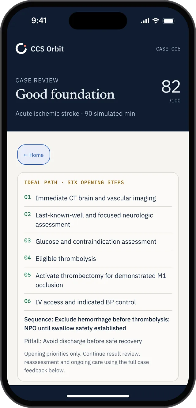
The sample stroke review includes immediate CT brain and vascular imaging among its opening priorities. Read the full case context when reviewing decisions.
Frequently asked questions
Why is the first CT for suspected stroke usually noncontrast?
The immediate question is whether intracranial hemorrhage is present. Further imaging, including CTA, depends on the stroke pathway and suspected vessel involvement.
Does every abdominal CT need IV contrast?
No. IV contrast is often used for suspected appendicitis or diverticulitis; suspected urinary stones commonly call for a noncontrast protocol. Acute mesenteric ischemia calls for a dedicated CTA protocol.
Is CT chest with IV contrast the same as CTPA?
Specify CT pulmonary angiography for suspected PE. CTPA uses a dedicated contrast-timed protocol to evaluate the pulmonary arteries.
Sources
- American College of Radiology, Cerebrovascular Disease and Head Trauma.
- American College of Radiology, Right Lower Quadrant Pain—Suspected Appendicitis and Left Lower Quadrant Pain—Suspected Diverticulitis.
- American College of Radiology, Imaging of Mesenteric Ischemia.
- American College of Radiology, Acute Onset Flank Pain—Suspicion of Stone Disease.
- American College of Radiology, Suspected Pulmonary Embolism.
For USMLE Step 3 CCS education only. This article is not medical advice. Actual imaging choices depend on the patient, current guidance, clinical judgment, and local protocols.